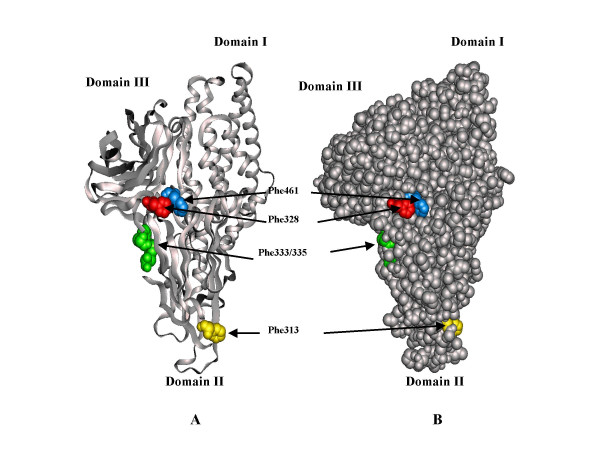
Figure 1.
Structure of Cry1Aa insecticidal protein showing surface phenylalanine residues (red) which were changed to alanine. A, ribbon diagram model to illustrate the secondary structure of domains I (alpha helical), II (beta strands) and III (beta sandwich). B, space filling model, to illustrate the surface exposure of the phenylalanine residues (red). Molecular models were generated using QUANTA (Molecular Simulations, Inc.)

