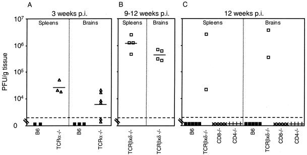
FIG. 4.
Replication of MAV-1 in T-cell-deficient mice. (A) Quantitation of virus from spleens and brains obtained at 21 days p.i. from B6 (▪) and TCRα−/− (▵) mice infected with 100 PFU of MAV-1. (B) Quantitation of virus from spleens and brains obtained at 9 to 12 weeks p.i. from moribund TCRβxδ−/− mice infected with 700 PFU of MAV-1. (C) Quantitation of virus from spleens and brains obtained at 12 weeks p.i. from B6 (▪), TCRβxδ−/− (□), CD8−/− (×), and CD4−/− (+) mice infected with 700 PFU of MAV-1. Virus levels were determined and are depicted as described in the legend to Fig. 1. Data points below the limit of detection were excluded in calculating mean titers and t statistics.

