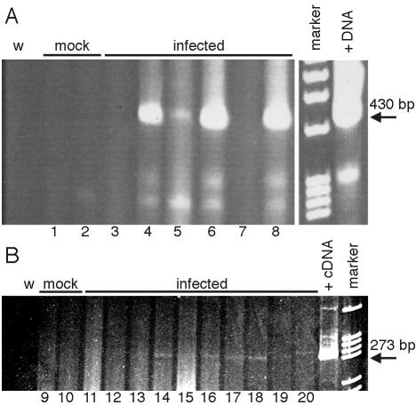
FIG. 5.
Presence of MAV-1 nucleic acid in B6 mice at 12 weeks p.i. (A) DNA was isolated from spleens (odd-numbered lanes) and brains (even-numbered lanes) of B6 mice mock infected or infected with 100 PFU of MAV-1 and was analyzed by PCR with MAV-1 E3-specific primers MAVR24718 and MAVR25148 for 55 cycles. The positive DNA control template (+DNA) was 1 μg of DNA isolated from an acutely infected mouse brain known to be positive for infectious virus, and the negative control was water (w). The arrow indicates the 426-bp PCR product from viral DNA. (B) RNA was isolated from spleens (odd-numbered lanes) and brains (even-numbered lanes) of B6 mice mock infected or infected with 700 PFU of MAV-1 and analyzed by RT-PCR with MAV-1 E3-specific primers MAVR24718 and MAVR25148 for 35 cycles. The positive cDNA control template (+cDNA) was from RNA isolated at 20 h p.i. from mouse 3T6 fibroblasts infected with MAV-1 at a multiplicity of infection of 5, and the negative control was water (w). The arrow indicates the 269-bp PCR product from viral cDNA. Each spleen and brain pair (lanes 1 and 2, 3 and 4, etc.) corresponds to a single animal.

