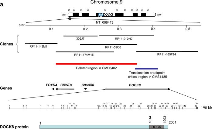
Fig. 1.
Mapping of the subtelomeric 9p chromosomal rearrangements. (a) Ideogram of chromosome 9 with a partial physical map of the subtelomeric 9p24.3 region, BAC clones used in FISH analysis, and known genes in italics are shown. Black arrows indicate the direction of the transcription. FISH analyses determined a critical deletion region (red bar) in patient CMS6482 and a region (blue bar) harboring the 9p24 translocation breakpoint in CMS1495. The genomic structure of the DOCK8 gene and a schematic of the 2031 amino acids DOCK8 variant are shown. The predicted DOCK domain in the C-terminal region is indicated. (b-d) FISH analyses in CMS6482. (b) Chromosome 9 subtelomeric probes (9p, green; 9q red) produced a green signal only on one chromosome 9 but not the other, indicating a 9p subtelomeric deletion in CMS6482. Red signal was present on both the normal and the derivative chromosomes 9. (c) RP11−59O6 (red) gave a signal (dark red) only on the normal chromosome 9 and not on the derivative chromosome 9 (arrow) indicating deletion of this probe in CMS6482. The chromosome 9 centromeric probe is in light red color. (d) Probe RP11−910H2 (green, arrowheads) is partially deleted on the derivative chromosome 9 and gave signals on both the normal and the derivative chromosome 9. The chromosome 9 centromeric probe is in light red color. (e) FISH analysis in CMS 1485. Probe RP11−910H2 (green) is distal to the 9p24.3 translocation breakpoint in CMS1485. The probe is present on the normal chromosome 9 as well as the derivative X chromosome. (f) PFGE analysis with a probe located in the 3’ portion of DOCK8 reveals an abnormally migrating fragment in CMS1485 but not in the control sample. The chromosome 9 centromeric probe is in light red color. (g) Fine mapping of the 9p critical region with quantitative genomic PCR in CMS6482, CMS1485 and in three controls (CMS11195, CMS5865 and CMS6265) reveals the 9p subtelomeric deletion in CMS6482 extends at least through exon 23 of the DOCK8 gene. CMS1485 has a deletion presumably associated with the translocation located between exon 23 and 34 with exon 29 deleted. The values represent the average of three replicates used in each case.


