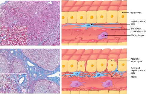
Fig. 1.
Azan-Mallory staining of human samples from normal (top) and cirrhotic (bottom) livers. Blue-stained area represents collagen and reticular fibers. Regenerative nodules surrounded by abundant fibrous band are observed in the cirrhotic liver (bottom). Schematic changes in the hepatic architecture between normal (right top) and cirrhotic (right bottom) livers are also shown.

