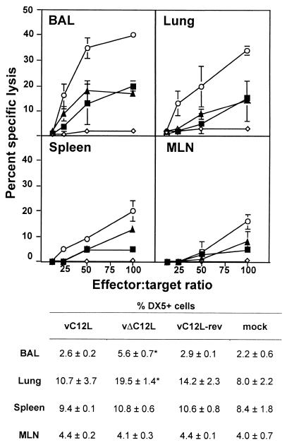
FIG. 3.
NK cytotoxicity in BAL, lung, spleen, and MLN after VV infection. Groups of six mice were mock infected (⋄) or infected with 104 PFU of vC12L (▪), vΔC12L (○), or vC12L-rev (▴). At day 3, NK cytotoxicity was determined in cell suspensions prepared from the BAL, lung, spleen, and MLN of mice. Specific lysis of YAC-1 cells was assessed by 51Cr-release assay. Data are expressed as the mean ± standard error from two groups of mice (n = 3 per group). Cells were also stained for expression of the pan-NK cell marker DX5 and examined by flow cytometry with a minimum of 20,000 cells analyzed in a lymphocyte gate. Results are expressed as the percentage of DX5+ cells in the total viable cell population. P values were determined by Student's t test and indicate the mean percentage of DX5+ cells from mice infected with vΔC12L that were significantly different from those from mice infected with vC12L or vC12L-rev. *, P < 0.05.

