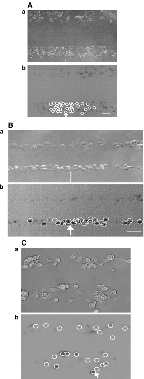
Fig. 4.
Propagation of a Ca2 + wave occurs between microglial lanes if these are not separated by distances greater than about 60  . A and B show parallel lanes of microglia in which the lane widths are 75±3
  and 25±3
  , respectively, separated by cell-free lanes of 165±3
  and 70±4
  , respectively; the open circles indicate the microglia that gave a Ca2 + response following mechanical excitation of the microglial cell indicated by the arrow; there is no propagation of the Ca2 + wave across these lanes. C shows parallel lanes of microglia in which lane widths are 50±6
  and the cell-free lane 46±10
  ; the open circles indicate that a Ca2 + wave response was able to propagate across cell-free lanes as well as along the lanes. The calibration bar is 45
  in A, B and C. The position of the micropipette in C(a) is not evident as it is out of focus

