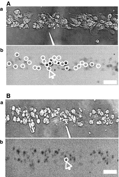
Fig. 5.
Propagation of Ca2 + waves is blocked by the ATP-degrading enzyme apyrase. A and B show the extent of propagation of Ca2 + , from the point of mechanical stimulation of a microglial cell, to other microglial cells in a lane. In each case, the top panel (a) shows the position of the mechanically stimulating micropipette and the lower panel (b) shows the microglial cells that gave a Ca2 + signal (open circles) in response to mechanical stimulation at the arrow. A is the control and in B apyrase (60 units/mℓ; grade III, Sigma) was present with only the stimulated cell now giving a Ca2 + transient. The calibration bar is 45

