
Figure 2. Intranuclear distribution of CFP-AR and YFP-RIP140.
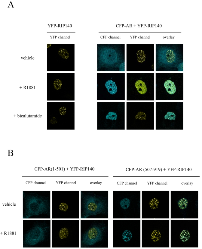
COS7 cells were transiently transfected with vectors expressing either the fusion protein YFP-RIP140 alone or together with CFP-AR (A), CFP-AR(1-501) (B, left panel) or CFP-AR(507-919) (B, right panel). Cells were incubated overnight in the presence of either vehicle or ligands (10−8 M R1881 or 10−6 M bicalutamide), fixed with 4 % paraformaldehyde and then observed using a Leica SP2 Confocal microscope. Pseudocolors were used (blue for CFP and yellow for YFP).