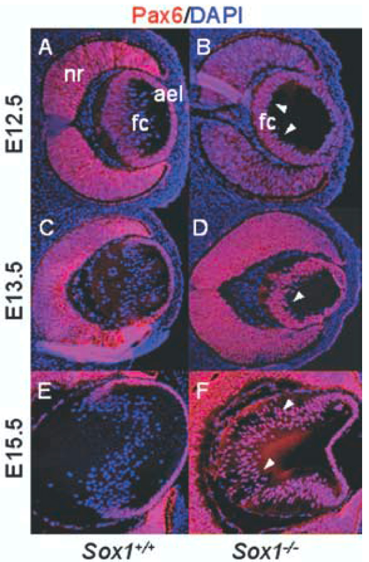
Fig. 2.

Pax6 expression is inappropriately maintained in the fiber cell compartment of Sox1−/− lenses. Wild-type (A, C, E) and Sox1−/− lenses (B, D, F) are shown with Pax6 immunofluorescence (red) and DAPI nuclear stain (blue) at E12.5 (A–B), E13.5 (C–D), and E15.5 (E–F). Arrowheads indicate fiber cell nuclei positive for Pax6.
