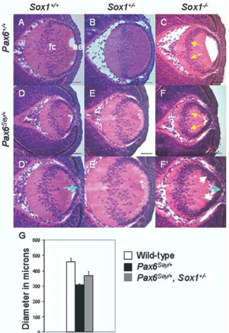
Fig. 4.

Sox1 heterozygosity partially rescues the size of Pax6Sey/+ lenses. Representative lenses at E13.5 are shown for wild-type (A); Sox1+/− (B); Sox1−/− (C); Pax6Sey/+ (D–D’); Pax6Sey/+, Sox1+/− (E–E’); and Pax6Sey/+, Sox1−/− (F–F’) lenses. High magnification images (D’–F’) show epithelial layer abnormalities. Yellow arrows indicate fiber cell nuclei that have remained in the lens posterior (C, F). White arrows indicate multi-layered AEL (F’) and blue arrows indicate pointed AEL (D’, F’). (G) Maximum lens diameters for wild-type (white box), Pax6Sey/+ (black box), and Pax6Sey/+, Sox1+/− (gray box) lenses are statistically different (see Experimental Procedures).
