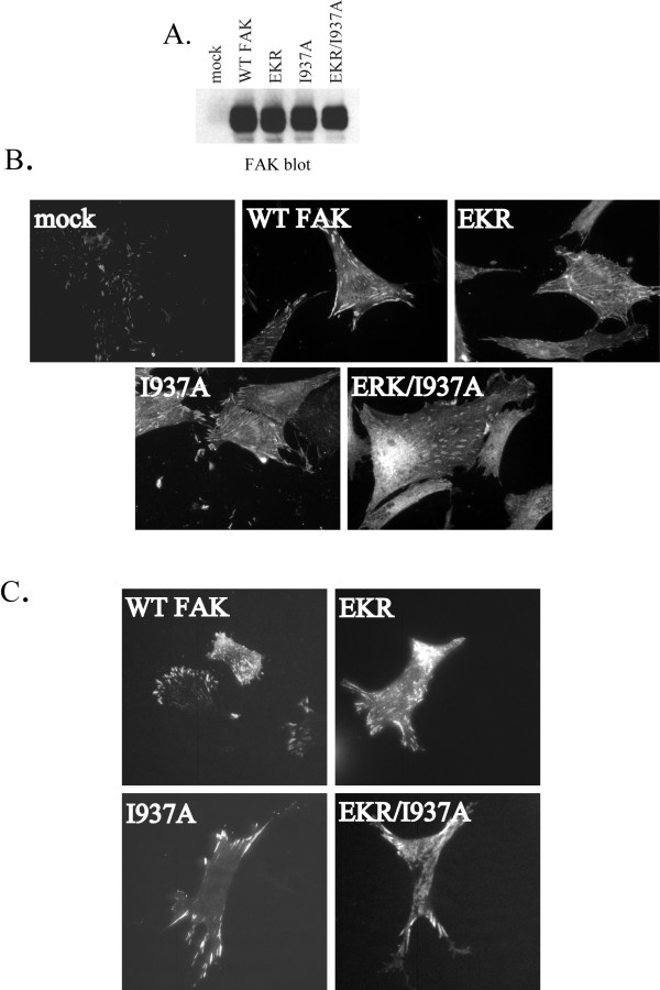
Figure 2.
Subcellular localization of FAK mutants. A) CE cells transfected with empty RCAS vector (mock) or RCAS constructs encoding wild type FAK or FAK mutants were lysed and 25 μg of lysate was blotted for FAK expression with BC4 antibody. B) CE cells transfected with empty RCAS vector (mock) or RCAS encoding wild type FAK or FAK mutants were plated on fibronectin-coated coverslips overnight, then fixed and used for immunofluorescent imaging using the FAK BC4 antibody. C) CE cells transfected with GFP-fusion constructs of wild type FAK or mutants were visualized in live cells by TIRF microscopy.

