Figure 6.
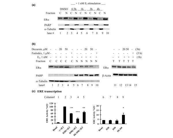
Effects of pyranocoumarins on estrogen receptor alpha nuclear translocation and transactivation in MCF-7 cells. (a), (b) Inhibitory effects of decursin on estrogen receptor (ER)α nuclear translocation in MCF-7 cells. (a) The time course of ERα nuclear translocation in MCF-7 cells stimulated by 17β-estradiol (E2) was first established in estrogen-starved MCF-7 cells. Fresh medium without or with 1 nM E2 was added. At different time points, nuclear (N) and cytosolic (C) fractions were prepared for western blot analyses. Poly(ADP-ribose) polymerase (PARP) and α-tubulin were detected as markers of N and C proteins, respectively. DMSO, dimethylsulfoxide. (b) For the decursin experiment, estrogen-deprived MCF-7 cells were pretreated with decursin for 2 hours and were stimulated with 1 nM E2 for 1 hour (total decursin exposure time, 3 hours). N and C fractions were prepared for western blot analyses. One-tenth nuclear fraction was used compared with the time-course experiment to show the difference. The total lysate ERα level was determined (right panel). (c) Differential effects of decursin versus decursinol on estrogen-response element (ERE) activity in MCF-7 cells: left, with E2; right, without, E2. An aliquot of 1 × 10⁵ MCF-7 cells was placed in a 12-well plate and cotransfected with ERE-luciferase reporter plasmid and pSV-β-galactosidase reporter in phenol-red free improved minimum essential medium (PRF-IMEM) without serum and insulin. After transfection for 24 hours, the cells were treated with different concentrations of decursin (D, 20 μM, 50 μM), decursinol (DL, 100 μM) or DMSO in the absence or presence of 1 nM E2 for 24 hours. Cells extracts were prepared for luciferase activity and β-galactosidase activity. Mean ± standard error, three independent experiments. *P < 0.05, **P < 0.01, ***P < 0.001 versus basal or E2-stimulated control.

