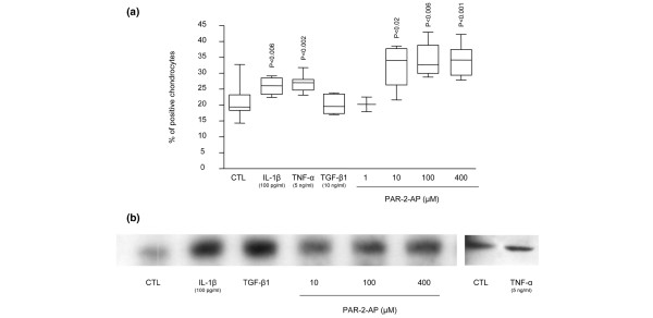
Figure 2.
Proteinase-activated receptor 2 (PAR-2) synthesis regulation. (a) PAR-2 immunostaining in osteoarthritis (OA) cartilage explants untreated (n = 16) and treated with interleukin 1 beta (IL-1β) (n = 9), tumor necrosis factor-alpha (TNF-α) (n = 9), transforming growth factor-beta-1 (TGF-β1) (n = 5), PAR-2-activating peptide (PAR-2-AP) 1 μM (n = 3), PAR-2-AP 10 μM (n = 4), PAR-2-AP 100 μM (n = 4), and PAR-2-AP 400 μM (n = 8) for 48 hours in Dulbecco's modified Eagle's medium (DMEM) 10% fetal calf serum (FCS). (b) Representative Western blot of PAR-2 synthesis in OA monolayer chondrocytes (n = 3) incubated for 72 hours in DMEM 2.5% FCS in the absence (CTL) or presence of IL-1β, TGF-β1, PAR-2-AP 10 μM, PAR-2-AP 100 μM, and PAR-2-AP 400 μM. P values indicate the comparison with the untreated (CTL) specimens.

