
Figure 3.
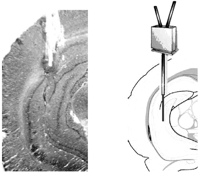
(Left.) Example of an acceptable cannula placement in which the active portion of the probe was positioned entirely within the hippocampus. (Right.) Schematic of a probe scaled to size overlaying a brain atlas section adapted from Franklin and Paxinos (1997).