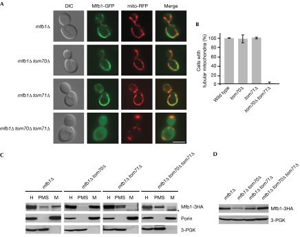
Figure 2.
Mfb1 mitochondrial targeting is disrupted in cells lacking Tom70 and Tom71. (A) Mfb1-GFP localization and mitochondria visualized by mito-RFP in mfb1Δ, mfb1Δ tom70Δ, mfb1Δ tom71Δ and mfb1Δ tom70Δ tom71Δ cells. Scale bar: 5 μm. (B) Mitochondrial morphology in wild-type, tom70Δ, tom71Δ and tom70Δ tom71Δ cells expressing mito-GFP. (C) Subcellular fractionation of strains used in (A) expressing Mfb1-3HA. The cell homogenate (H) was separated into post-mitochondrial supernatant (PMS) and mitochondrial pellet (M). Porin and 3-PGK were monitored as mitochondria and cytoplasm markers, respectively. The asterisk indicates nonspecific bands. (D) Steady-state levels of Mfb1-3HA in total cell extracts of strains used in (C). 3-PGK was monitored as a loading control. DIC, differential interference contrast; GFP, green fluorescent protein; mito-RFP, mitochondria-targetted red fluorescent protein; PGK, phosphoglycerate kinase; TOM, translocase of the outer membrane.

