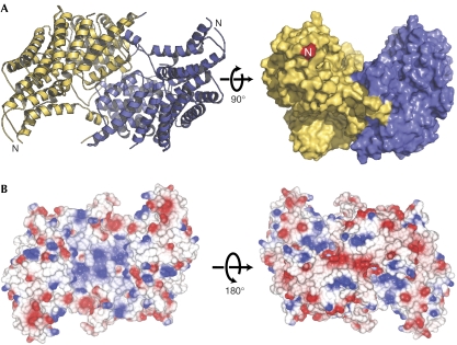
Figure 3.
Structure of the Cep3Δ dimer and electrostatic properties. (A) The proposed biological dimer, formed around a crystallographic twofold axis. A space filling view, rotated by 90° shows the groove on the surface. (B) Electrostatic surface representations of the dimer on the concave (left) and convex (right) surfaces.

