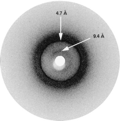
Figure 2.
X-ray diffraction pattern of PI3-SH3 fibrils. The reflections generated by the interstrand spacing (4.7 Å) and the intersheet spacing (centered at 9.4 Å) typical of amyloid fibrils are marked by arrows. Because of poor alignment of the fibrils, these reflections appear as rings and do not display a meridional or equatorial character.

