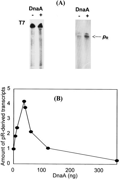
Figure 2.
Activation of the pR promoter by DnaA protein in vitro. (A) Examples of autoradiograms after in vitro transcription experiments. (Left) In the control, 0.5 μg of bacteriophage T7 DNA was used as a template. (Right) In the experiment, 0.5 μg of a HindIII–DraI fragment of plasmid pKB2 was used as a template DNA. The reaction mixtures contained no DnaA (lanes −) or 35 ng (final concentration, 26.6 nM) of DnaA (lanes +). The position of pR-derived transcripts (812 nucleotides long) is indicated. (B) Average data from six experiments of in vitro transcription. Template DNA (0.5 μg) and DnaA protein (as indicated) were used, and appropriate bands on the autoradiograms were quantified by densitometry. Results very similar to those obtained with the HindIII–DraI fragment of pKB2 were observed when NsiI–NsiI fragment of pKB2 or whole pKB2 plasmid linearized with BstXI were used as DNA templates (the lengths of pR-derived transcripts were 288 and 269 nucleotides, respectively). The positions of appropriate restriction sites in λ plasmid DNA are depicted in Fig. 1.

