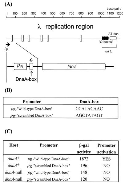
Figure 3.
(A Upper) Map of the replication region of bacteriophage λ (also present in λ plasmids) from the pR promoter to oriλ. The scale is given in base pairs, the pR promoter is marked by the thick arrow, putative DnaA boxes are represented by vertical rectangles, and sequences recognized by the O replication initiator (O boxes) and the A+T-rich region are also indicated. (A Lower) The fragment of λ DNA present in the lacZ fusions is indicated, and the orientation of the DnaA-box is marked by arrow (this part of the figure is not drawn to scale). (B) Sequences of the wild-type and scrambled DnaA boxes present in appropriate fusions. (C) The activity of β-galactosidase per single copy of the fusion is presented (in a table form) for both fusions in dnaA+ and dnaA-null hosts. It seems that pR is activated only in the wild-type host harboring the “wild-type” fusion (YES in the table) and that the lack of either functional DnaA protein or DnaA-binding site immediately downstream of pR results in no activation of the promoter (NO in the table), and the obtained values reflect residual pR activity.

