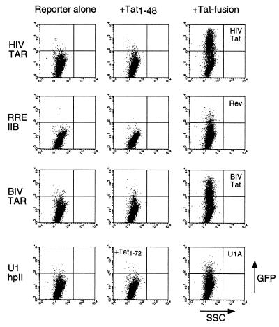
Figure 2.
Activation of GFP expression by Tat fusion proteins and corresponding RNA reporters. Cells were lipofected with HIV TAR, RRE IIB, BIV TAR, or U1 hairpin II GFP reporters (2 μg) alone (Left), along with Tat1–48 or Tat1–72 (Center), or along with full-length HIV Tat or Tat fusions to a Rev peptide, BIV Tat peptide, or U1A RNA-binding domain, respectively (4 μg) (Right). Rev and BIV Tat peptides were fused to Tat1–48 whereas the U1A domain was fused to Tat1–72 to ensure nuclear localization. Plots show relative GFP fluorescence on the y axis and relative side scatter (a measure of cell granularity) on the x axis for 10,000 cells.

