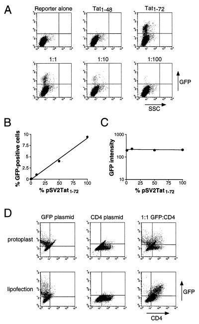
Figure 3.
Plasmid delivery by protoplast fusion. (A) FACS analysis of cells containing a stably integrated HIV LTR-GFP reporter (reporter alone) or reporter cells fused with protoplasts containing pSV2Tat1–48 or pSV2Tat1–72 plasmids, or fused with protoplast mixtures containing pSV2Tat1–72 and pSV2Tat1–48 in 1:1, 1:10, or 1:100 ratios. (B) Plot of the percentage of GFP-expressing cells as a function of the proportion of pSV2Tat1–72 in the mixture, from A. Based on the percentage of positive cells obtained with pSV2Tat1–72 alone, we estimate that ≈10% of cells fused productively in this experiment. (C) Plot of GFP intensity as a function of the proportion of pSV2Tat1–72 in the mixture, from A. (D) Protoplasts were prepared containing CMV-GFP or CMV-CD4 expression plasmids and were fused to HeLa cells either separately or in a 1:1 mixture, and analyzed for protein expression by FACS (Upper). Plasmid DNAs also were introduced by lipofection and analyzed (Lower).

