
Fig. 2.
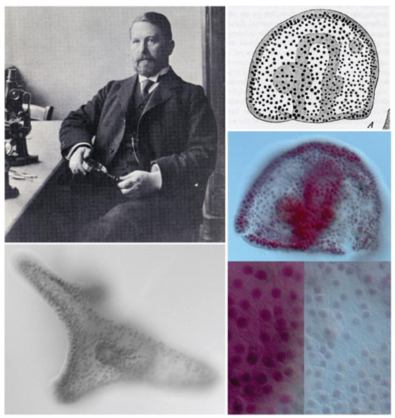
Provided by Dr. Andrew Ransick. Boveri at his laboratory at the Institute of Zoology in Würzburg in 1907, reproduced from Baltzer, 1967. Boveri’s drawing at upper right shows an abnormal aneuploid gastrula stage embryo containing nuclei of two different sizes. Below is the same embryo, photographed by Dr. Andrew Ransick from one of Boveri’s original 1904 slides, which had been kindly provided to the author by Professor Ulrich Scheer, University of Würzburg. In the magnification below the smaller nuclei in the blue region to the right can clearly be distinguished. Lower left shows an approximately normal pluteus larva from the same series of experiments.