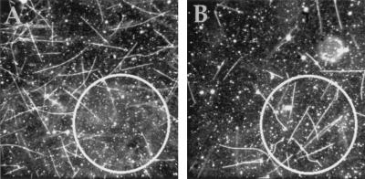
Figure 2.
CALI of HAkinesin using fluorescein-labeled anti-HA Fabs in motility assays. (A) An area with a diameter of 75 μm (indicated by a circle) was illuminated through the fluorescein filter set in the absence of microtubules (only immobilized HAkinesin and HA-kinesin-bound, fluorescein-labeled Fab fragments were present). Ten minutes after adding the microtubules and washing the flow cell (see Materials and Methods), microtubules were observed. (B) As in A, but the area was illuminated after adding the microtubules. (Because some gliding microtubules detach from the surface after washing the flow cell, but hardly any immobilized ones do so, the density of microtubules is higher in the illuminated area of B. In experiment B a lower concentration of microtubules was used than in A. Typical microtubule densities are in Table 2.)

