
Figure 5.
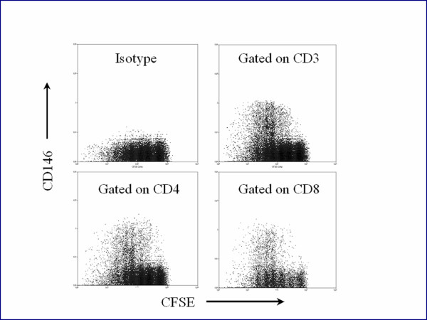
Mononuclear cells from the peripheral blood of a normal volunteer were incubated with carboxyfluorescein diacetatesuccinimidylester (CFSE) and PHA, as described in the text. CFSE fluorescence reduced with each cellular division. The cells were cultured a total of five days. The upper left panel displays CFSE staining versus a PE isotype control of CD3+ cells. The upper right panel shows staining of PECD146 versus CFSE of CD3+ T cells. Lower panels display CFSE vs CD4 and CD8. CFSE staining decreased with each successive division of the T cells, and CD146 expression increased.