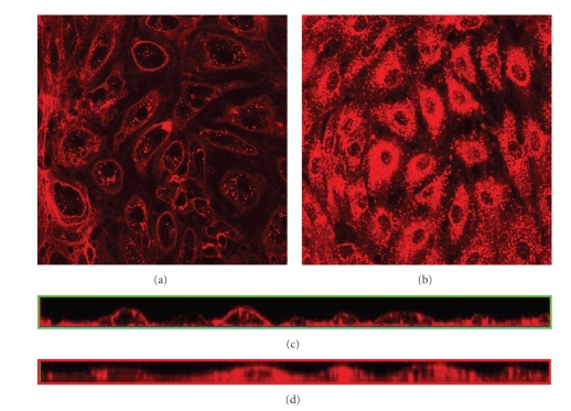
Figure 3.
Effects of PAO on internalization of FITC-conjugated lectins. The GL is visualized under the confocal microscope through staining of heparan sulfate with Texas Red-conjugated lectin after 24 hours in culture. (a) Cells grown in static condition, 45 minutes prior to staining medium were changed to EMDM with 10 μM Phenyl Arsine Oxide to prevent endocytosis, (b) cells grown under static condition. Images are cross-sections of the cell monolayer = 0.5–2 μm with = 0 at the glass surface, (c) cross-sections from the side of the stack of images of (a) created with confocal microscopy, and (d) cross-sections from the side of the stack of images of (b) created with confocal microscopy.

