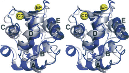
Figure 3.
Stereoview of the superimposed structures of Ca2+-free (silver) and Ca2+-bound (blue) rat α-PV. The two structures were superimposed so as to minimize the overall RMSD. Coordinates for the Ca2+-bound structure were obtained from PDB 1RWY (Bottoms et al. 2004).

