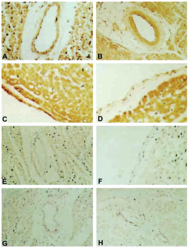
FIG. 11.

Evidence for nitrotyrosine formation in human myocardial inflammation. Representative examples are shown of nitrotyrosine immunoreactivity in cardiac tissue samples from patients with myocarditis, sepsis, or no cardiac disease (control patients). A: low-power (×20) photomicrograph of nitrotyrosine immunoreactivity (brown staining) in the myocardium, vascular endothelium, and vascular smooth muscle of myocarditis patient. B: low-power (×20) photomicrograph of nitrotyrosine immunoreactivity in the myocardium, vascular endothelium, and vascular smooth muscle of sepsis patient. Note the relative absence of staining of the connective tissue elements. C: higher-power (×40) photomicrograph of intense nitrotyrosine immunoreactivity in the endocardium of myocarditis patient. D: higher-power (×40) photomicrograph of nitrotyrosine immunoreactivity in the endocardium of sepsis patient. E: low-power (×20) photomicrograph of minimal nitrotyrosine immunoreactivity in the myocardium and the virtual absence of nitrotyrosine immunoreactivity in the vascular endothelium and vascular smooth muscle of control patient 1C. F: higher-power (×40) photomicrograph of nitrotyrosine immunoreactivity in the endocardium of control patient. G: low-power (×20) photomicrograph demonstrating the inhibition of nitrotyrosine immunoreactivity by the preincubation of the primary antibody with 10 mM nitrotyrosine before tissue staining in myocarditis patient. H: low-power (×20) photomicrograph demonstrating the inhibition of nitrotyrosine immunoreactivity by the preincubation of the primary antibody with 10 mM nitrotyrosine before tissue staining in sepsis patient. [From Kooy et al. (707) with permission from Lippincott Williams & Wilkins.]
