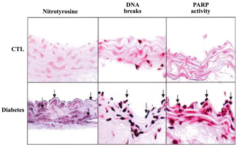
FIG. 18.

Evidence for nitrotyrosine formation and PARP activation in diabetic vasculature. Immunohistochemical staining for nitrotyrosine, terminal deoxyribonucleotidyl transferase-mediated dUTP nick-end labeling (indicator of DNA breaks), and poly-(ADP-ribose) (index of PARP activity) in control rings (top row) and in rings from diabetic mice (bottom row). [Derived from Garcia Soriano et al. (430) with permission from Nature Publishing Group.]
