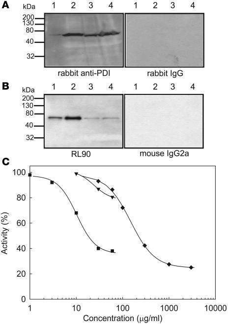
Figure 1. Interaction of anti-PDI antibodies with human and mouse PDI.
Lysates of human (1 × 10⁹ platelets/ml) and mouse (1 × 10¹⁰ platelets/ml) platelets were subjected to SDS gel electrophoresis followed by immunoblotting with anti-PDI antibodies. (A) Polyclonal anti-PDI antibodies. Lane 1, recombinant human PDI (20 ng); lane 2, human platelet lysate (20 μl); lane 3, mouse platelet lysate (5 μl); lane 4, mouse platelet lysate (20 μl). Left: Immunoblot developed using affinity-purified rabbit anti-bovine PDI antibody. Right: Immunoblot developed with control nonimmune IgG. (B) Monoclonal antibody RL90 to PDI. Lane 1, recombinant human PDI (1 ng); lane 2, human platelet lysate (1 μl); lane 3, mouse platelet lysate (5 μl); lane 4, mouse platelet lysate (20 μl). Left: Immunoblot developed using the RL90 anti-PDI monoclonal antibody. Right: Immunoblot developed with control IgG2a monoclonal antibody. (C) PDI activity was measured with the insulin transhydrogenase assay in the releasate of thrombin-activated mouse platelets (n = 3) in the presence or absence of 1–60 μg/ml RL90 monoclonal anti-PDI antibody (squares), 10–60 μg/ml rabbit anti-PDI antibody (inverted triangles), or 0.03–3.0 mg/ml bacitracin A (diamonds). Results are shown as a mean of 2 experiments.

