
FIGURE 1.
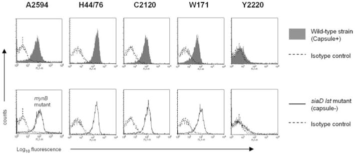
fH binding to N. meningitidis. A representative strain of N. meningitidis from each of the five major pathogenic serogroups (A–C, W-135, and Y) was chosen for study. Bacteria (~108 organisms) were incubated with 2 μg of pure fH, and bound fH was detected by flow cytometry using goat anti-human fH Ab. Upper panel, fH binding to encapsulated wild-type strains (shaded histogram). Lower panel, fH binding to unencapsulated mutant derivatives that also lacked the ability to sialylate their LOS (mynB mutant of A2594 and siaD lst mutants of the remaining four strains; solid line). Isotype controls (no fH added) are indicated by the broken lines. The x-axis represents fluorescence in arbitrary units on a log10 scale, and the y-axis represents the number of events.