
Fig. 6.
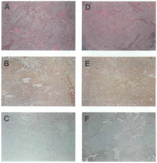
Immunohistochemical staining of sections of melanoma in situ. Sections from tumor specimens described in Table 1 from patient Nos. 1106 (panels A-C) and 1180 (panels D-F) are shown. Immunohistochemical staining in panels B, C, E, and F was done at the same time, under exactly the same conditions with anti-human beta2-microglobulin (β2m) monoclonal antibody followed by immunoperoxidase staining (see “Materials and Methods” section for details). Strongly positive and weakly positive staining for β2m is shown in panels B and E, respectively. Panels C and F are examples of specimens that stain negative for β2m. Hematoxylin–eosin stains are shown in panels A and D, where melanin deposits that appear dark brown can be seen.