
Figure 1.
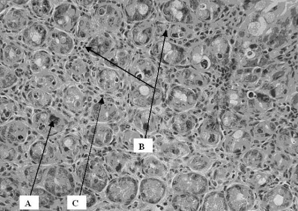
Cross section of intestinal wall 48 hrs after 19 Gy irradiation. An increase in apoptosis (A), intraepithelial granulocytes (B) and lymphocytes (C) was observed. The slides were stained with hematoxylin and eosin for histological evaluation under light microscopy, which was done by the pathologist in a blinded fashion.