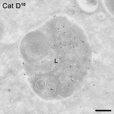
Fig. 5.
Immuno-EM of a B lymphoblast derived from a patient with I-cell disease showing a lysosome (L) positively labeled for the lysosomal hydrolase cathepsin D (represented by 10 nm gold particles). This picture indicates that although the MPR pathway is impaired in these cells, lysosomal enzymes can still reach lysosomes. Bar 200 nm

