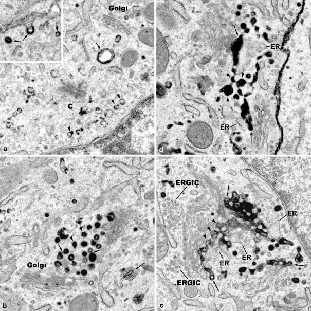
Fig. 1.
The Golgi apparatus reorganizes during endocytosis of WGA within a period of 30 min. ainsert: The insert shows WGA-HRP reaction products concentrated in a clathrin-coated pit, and a coated vesicle (arrows), as well as lining the membrane of a small uncoated vesicle (arrowhead). ×35,000. a Small endosomes (arrowheads), and a large vacuolar endosome (arrow) are apparent in the cytocentre (c), and close to a small Golgi apparatus stack (Golgi). A distinct TGN is not visible. ×30,000. b Globular early endosomes are accumulated close to the trans side of Golgi apparatus stacks (Golgi); some of the endosomes are covered with clathrin coats (arrowheads); partly, they exhibit WGA reaction products attached to the membranes, partly contained within the lumina. Some of endosomes appear to contact each other, and fine bridges are visible (short arrows). WGA reactions are apparent within the cisternae of a small Golgi apparatus stack (large arrow). ×31.500. c A network, an endocytic trans Golgi network (endocytic TGN), is apparent consisting of interconnected globular pieces (arrowheads) that resemble the earlier globular endosomes. Again here, the WGA reaction products are either attached to the membranes, or fill the lumina. Parts of this endocytic TGN are attached to stacks of Golgi cisternae at their trans sides (arrows), and are associated with trans Golgi ER. At the cis side, ER-Golgi-intermediate compartments (ERGIC) are visible. ×28,000. d Endocytic TGN consisting of interconnected globular pieces (arrowheads) and filled with WGA reaction products are partly attached to Golgi apparatus stacks (arrows), and associated with trans Golgi ER. ×32,000

