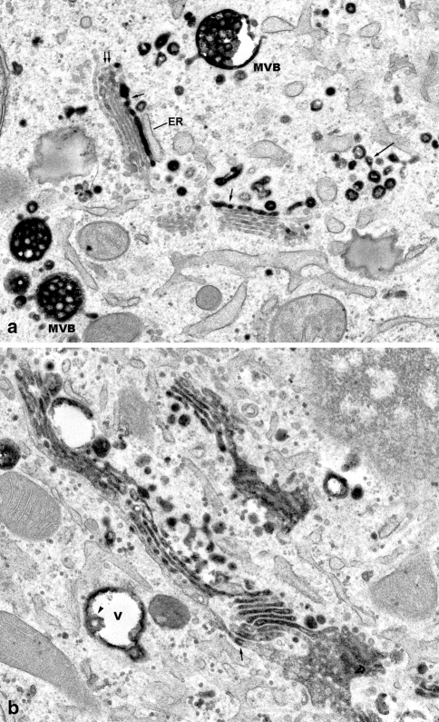
Fig. 4.
WGA is taken up into the stacked Golgi cisternae a Free portions (long arrows), and Golgi-integrated portions (short arrows) of the WGA-reactive endocytic TGN are visible. Fine WGA reactions also are apparent at confined regions of some of the stacked Golgi cisternae (double arrow). Several multivesiculated bodies (MVB) are densely filled with WGA-reaction products, and show domains, where tubular and vesicular transport carriers are formed. ×20,000. b A Golgi ribbon is visible, in which the individual stacks of cisternae are interconnected at the cis, as well as at the trans side. Most of the stacked Golgi cisternae are densely filled with WGA reaction products; others show reactions at limited regions (arrow). At the right lower corner, connections of medial cisternae with the cis-most cisterna of the same stack, and of other stacks are shown. In a vacuolar endosome (V), WGA reaction products are attached to the limiting membrane, and to an intra-vacuolar vesicle (arrowhead). ×23,000

