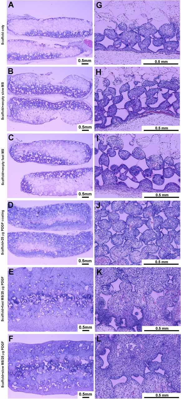
Figure 1. PDGF-containing microspheres (MS) in nanofibrous scaffolds (NFS) stimulates tissue invasion in vivo.
Left panel at low magnification (2X) shows the tissue completely penetrated the entire NFS for the groups with PDGF encapsulated in PLGA microspheres, while other groups show a portion of scaffold occupied by ingrown tissue. In addition, the scaffolds were enlarged by the penetrated tissues in the groups with high dose PDGF encapsulated in PLGA microspheres. Right panel at high magnification (10X) demonstrated that most of the cells in the penetrated tissues were fibroblast-like cells and lymphocyte-like cells. On the scaffold pore surfaces multiple macrophage-like cells were seen. Bars indicate 0.5 mm.

