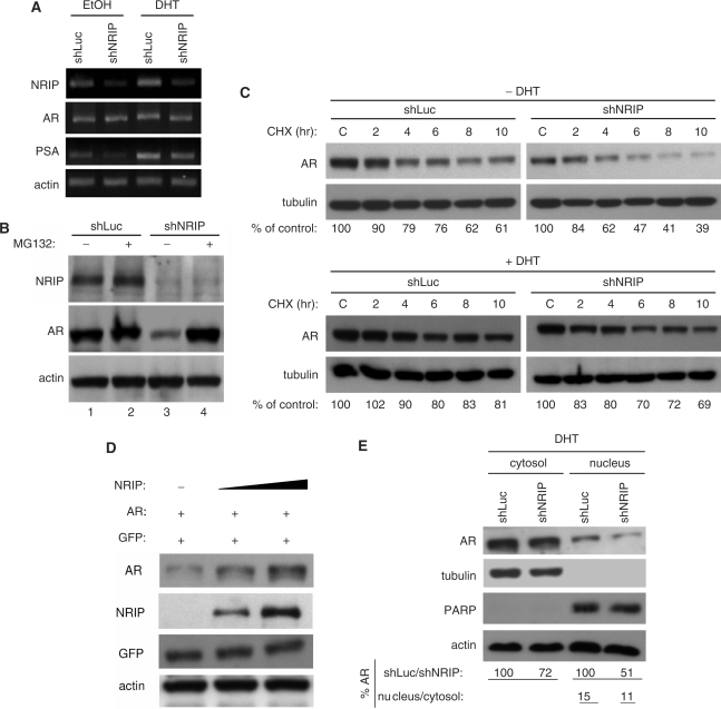
Figure 6.
NRIP stabilizes AR protein but has no effect on AR mRNA and AR nuclear translocation. (A) The effect of NRIP on AR mRNA. LNCaP cells were infected with LV-shNRIP (or LV-shLuc as control) in the presence or absence of DHT. Three days post-infection, total RNA was extracted and subjected to semi-quantitative RT-PCR using NRIP, AR, PSA or β-actin primers. (B) NRIP effects on proteasome-dependent AR protein degradation. Lentivirus encoding shNRIP infected LNCaP cells in the presence of DHT and with or without proteasome inhibitor (MG132) for 24 h. Cell lysates were analyzed and immunoblotted with anti-NRIP, AR and actin antibodies as indicated. (C) NRIP effects on AR protein stability. LNCaP cells were infected with LV-shNRIP or LV-shLuc. Three days after infection, cycloheximide (CHX) was added for the indicated time. The amounts of AR protein from the lysates of cells in the absence (upper panel) or presence (lower panel) of DHT were analyzed by western blotting using anti-AR and anti-tubulin (as a loading control) antibodies. The% of control indicates the AR amount at each time point relative to the control (without CHX treatment, set at 100). (D) AR stabilization by NRIP in a dose-dependent manner. 293T cells were co-transfected with 1 μg pcDNA3.0-AR, 0, 2 or 4 µg pNRIP-F0lag and 1 µg EGFP-C1 as transfection efficiency controls in a 6-well plate. Total plasmid DNAs were adjusted by pcDNA3.1. Cells were cultured in CDS medium. Forty-eight hours after transfection, cell lysates were subjected to western blotting using antibodies against AR, Flag (for NRIP), GFP and β-actin. (E) The subcellular location of AR by NRIP. LNCaP cells were infected with LV-shLuc and LV-shNRIP for 3 days in the presence of DHT. Cell lysates were separated by cytosol and nuclear fractionation and followed by western blot analysis. The expression level of AR protein was quantified by UVP imaging and normalized by tubulin for the cytosolic fraction and by PARP for the nuclear fraction. For AR stability analysis, proteins from shLuc-treated LNCaP cells were defined as 100%. The percentage of AR nuclear localization was calculated as cytosol/nucleus ratios in shLuc-treated or shNRIP-treated LNCaP cells.

