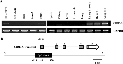
Figure 1.
Cell- and tissue-specific expression and genomic structure of the human CIDE-A gene. (A) RT-PCR analysis of CIDE-A expression in five human cell lines and eight human tissues as indicated. Five micrograms of total RNA from cells and tissues was analysed by RT-PCR using primers specific for CIDE-A CDS. A 305-bp fragment of CIDE-A CDS was amplified. A 532-bp fragment of the GAPDH gene was used as a positive control for RNA integrity. (B) Structure of the human CIDE-A locus. Boxed arrows represent published cDNAs encoding known proteins. The open, grey and black boxes represent the untranslated regions, ORFs and predicted CpG island, respectively.

