Figure 5.
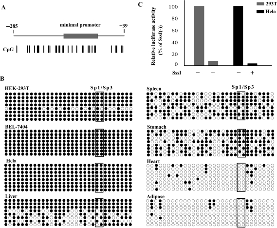
Methylation status of CpG sites in the CIDE-A promoter. (A) Schematic diagram of the CIDE-A gene 5′ region. Numbers indicate the positions relative to the translation start site of CIDE-A. The CIDE-A minimal promoter is indicated by the grey box. The position of the 31 CpG sites detected by bisulfite sequencing is indicated by a short vertical line. (B) Bisulfite sequencing of the CIDE-A promoter region. The genomic DNA from BEL-7404, Hela and HEK-293T cells and from the liver, spleen, stomach, heart and adipose tissues was treated with bisulfite and amplified by PCR. The PCR products were then subcloned and sequenced. For each cell line and tissue, the methylation status of 31 CpGs in the promoter region is shown for eight clones. White and black circles indicate unmethylated and methylated CpGs, respectively. Circles enclosed by boxes represent the CpGs in the Sp1/Sp3-binding sites. (C) Effect of in vitro methylation on the activity of the CIDE-A promoter. The CIDE-A promoter-luciferase plasmid p(−250/−13) was methylated in vitro by SssI methylase. Methylated and unmethylated p(−250/−13) were then transiently transfected into Hela and HEK-293T cells and assayed for luciferase activity. Results are shown as percentages of luciferase activity, with the activity due to unmethylated p(−250/−13) considered as 100%. The average ± SD values from three independent experiments performed in triplicates are shown.

