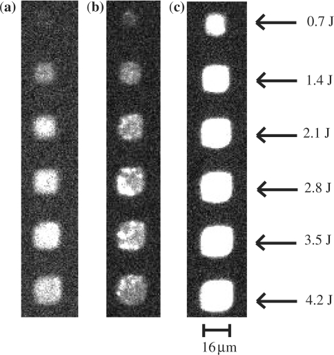
Figure 6.
Fluorescence microscope images of a Cy3-labeled complementary DNA sequence hybridized to identical probe sequence fabricated using an increasing UV dose (measured in Joules) on glass (a), glassy carbon (b) and diamond (c). Each feature is a single pixel (16 μm × 16 μm) separated by 16 μm. The fluorescence observed from the diamond substrate is greater than that from either glass or glassy carbon. This may be the result of the polished silicon acting as a mirror behind the thin film diamond.

