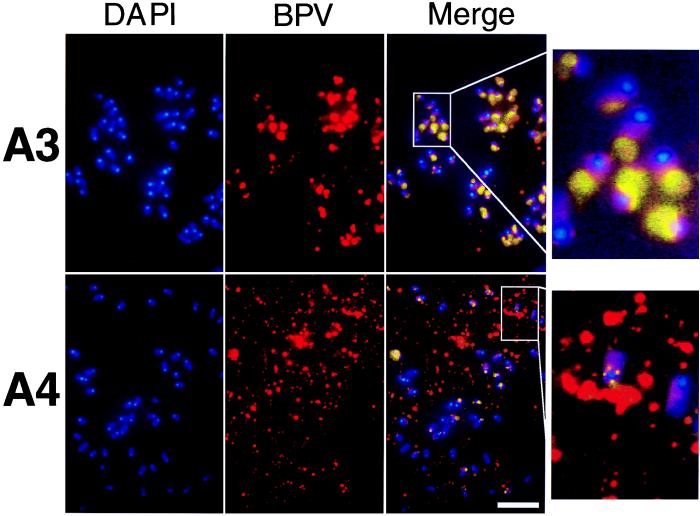
Figure 3.
Colocalization of BPV DNA with cellular metaphase chromosomes. DNA staining with DAPI revealed mouse chromosomes that showed predominantly acrocentric organization with brightly staining centromeric regions. BPV DNA was detected by FISH. The chromosomes from one mitotic plate are shown in each panel. Intensely colocalizing BPV and DAPI staining produced magenta, which was pseudocolored yellow, and lower-intensity colocalization produced pink. (Right) Magnifications ×4 of a small region of the merged images. The neor control background levels of staining were similar to those shown in Fig. 2A. (Bar = 5 μm.)

