Figure 5.
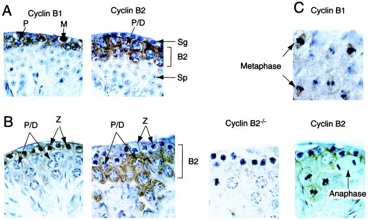
Cyclin B1 and B2 localization in the testis. Sections of testis, prepared from cyclin B2 nullizygous mice or their heterozygous littermates, were analyzed by immunostaining with the indicated antibodies (brown) and counterstained with hematoxylin (blue) The sections in A show mitotic precursors (M, Sg) and postmeiotic spermatocytes (Sp). Cyclin B2 staining is seen in late pachytene and diplotene cells, whereas cyclin B1 staining is most intense at the earlier zygotene stage. The postmeiotic spermatocytes do not stain with either anti-B type cyclin antibody. B represents stages X or XI of spermatogenesis, cell types are: P/D, late pachytene or diplotene; Z, zygotene. A section from a B2 nullizygous animal is shown as a control. C shows stage XII sections with fields of meiotic metaphases and an anaphase from the same series of testis sections (B2+/− heterozygotes), stained with anti-cyclin B1 (Upper) or anti-cyclin B2 (Lower). Note the disappearance of cyclin B2 at anaphase, and the association of both B-type cyclins with mitotic spindles. The upper two metaphase figures in the cyclin B1 panel probably represent meiosis I, and the lower set are probably meiosis II, judged by position in the section and the intensity of chromosome staining.

