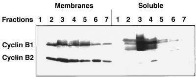
Figure 6.
Cyclin B1 and B2 differ in their subcellular localization. Mouse testes were homogenized in a Hepes-sucrose buffer and nuclei were removed by low speed centrifugation. The postnuclear extract was loaded on a 4 ml sucrose step gradient and centrifuged overnight in a Beckman SW55 rotor at 30,000 rpm (105 g). The fractions collected from the gradient were diluted and spun for 45 min in a Beckman TL100 benchtop ultracentrifuge. The membranous pellet was dissolved in SDS/PAGE sample buffer. The protein in the supernatant was precipitated with methanol and chloroform, dried, and dissolved in sample buffer. The fractions were analyzed by immunoblotting with anti-cyclin B1 and B2 antibodies.

