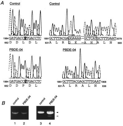
Figure 5.
Mutation analysis of PEX1 from a CG-I patient. (A) Nucleotide sequence analysis of PBDE-04 PEX1. Partial sequence and deduced amino acid sequence of PEX1 cDNA isolated from patient PBDE-04 and a normal control are shown. (Left) One-base point mutation (shaded), T to C in the codon for Leu-664, resulting in a codon for Pro-664 in one allele (see the open arrowhead in Fig. 2). (Right) A 171-bp deletion of nucleotide residues 1900–2070 (boxed), in another allele (solid arrowheads in Fig. 2). (B) RT-PCR analysis of PEX1 transcript from a control and PBDE-04. RT-PCR of poly(A)+ RNA was done with two sets of primers, RT1 and RT2 to amplify full-length PEX1 (lanes 1 and 2), and F5 and R6 to amplify a partial PEX1 fragment, nucleotide residues from 1276 to 2152 (lanes 3 and 4). Lanes: 1 and 3, a control; 2 and 4, PBDE-04. Note that two products with a normal size, ≈0.9 kb (solid arrowhead), and a smaller, ≈0.7 kb (open arrowhead), were obtained in PBDE-04 (lane 4).

