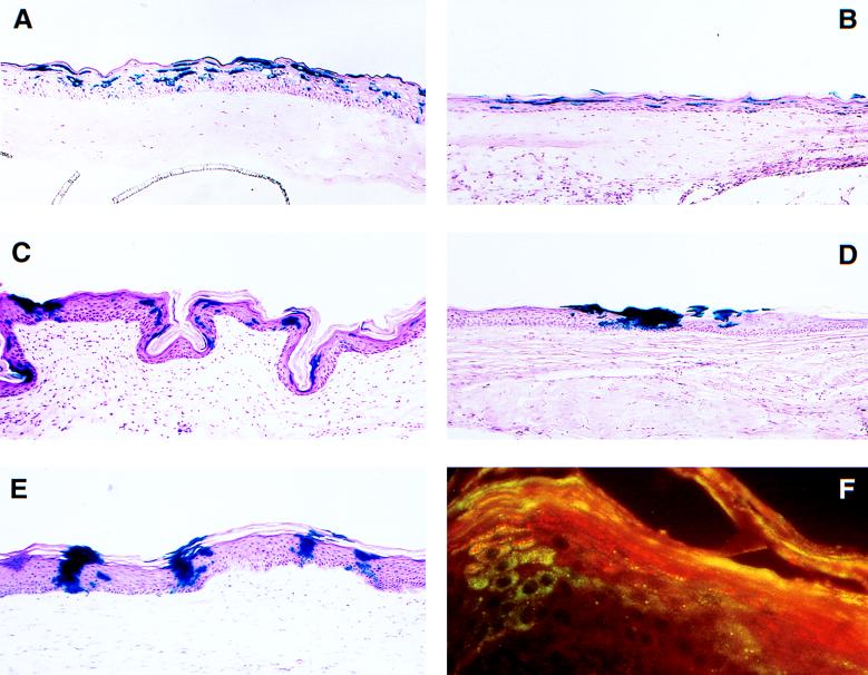
Figure 1.
Histochemical staining of grafted tissue for transgene expression. Organotypic raft cultures constructed with MFGlacZ-transduced keratinocytes were grafted to athymic mice. At the times noted, the tissue was excised and examined for β-gal expression by staining with X-Gal. β-gal positive cells are present in all grafts. (A) Organotypic raft culture before grafting. (B) One week after grafting. (C) Ten weeks after grafting. (D) Twenty weeks after grafting. (E) Forty weeks after grafting. (F) A 10-week graft stained with an antibody for β-gal (green) and an antibody for filaggrin (red). Note dual expression of β-gal and filaggrin (orange) in the column of cells on the left. (A–E, ×240.)

