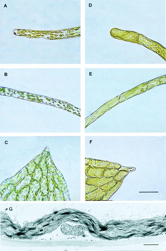
Figure 3.
Phenotypes of transgenic Physcomitrella patens generated by the construct outlined in Fig. 2C. Light microscopy from different tissues from transgenic plants with wild type-like plastids (A–C), and macrochloroplasts (D–F), respectively. The tissues depicted are chloronema (A and D), caulonema (B and E), and leaves (C and F). See ref. 30 for a review on moss development and tissue definition. Bar = 100 μm. As judged by transmission electron microscopy, division of chloroplasts was impeded in ΔPpftsZ knockouts, whereas form and number of mitochondria remained unchanged (G). (Bar = 1 μm.)

