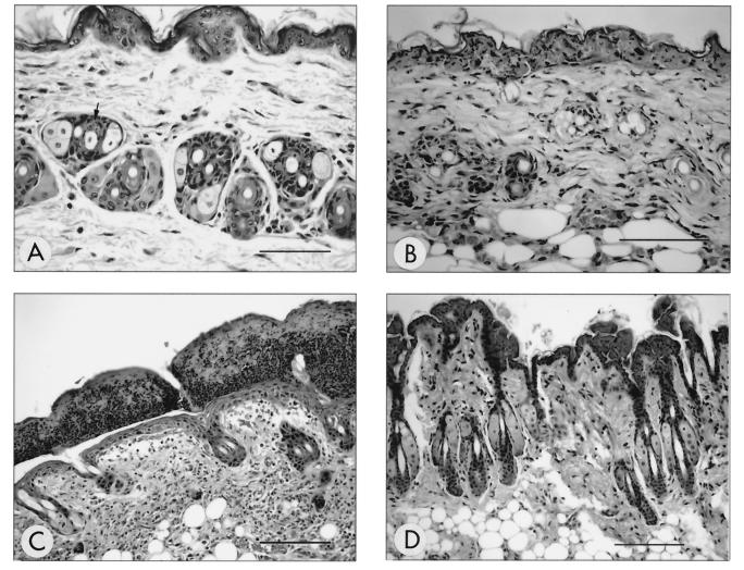
FIG. 1.
Temporal development of histological lesions in haired skin. Temporal development of skin lesions in mice inoculated with HVP2nv strains was characterized by subtle necrosis of hair follicle epithelium observed as early as 1 day p.i. (A) (arrow) followed by more conspicuous necrosis involving widespread adnexal structures by 5 days p.i. (B). By 9 days p.i. (C), the epidermis was overlaid by a thick serocellular crust and the dermis contained a primarily mononuclear inflammatory infiltrate (dermatitis) with evidence of follicular destruction and effacement. Cutaneous lesions were not observed at any time point following inoculation with HVP2ap (D) (9 days p.i.). All sections were H&E stained. Bar for panels A and B = 150 μm; bar for panels C and D = 40 μm.

