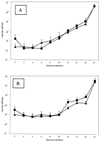
FIG. 4.
HVP2 in vitro growth kinetics. Vero cell monolayers were infected with 5 PFU of various HVP2 isolates representing the two HVP2 pathogenic subtypes/cell and incubated at 37°C for 48 h. Data points represent mean PFU values of four isolates from HVP2nv (▴) and HVP2ap (▪) at every time point for both intracellular virus (A) and virus present in the extracellular fluids (B). There was no statistical difference between the replication kinetics of HVP2ap and HVP2nv.

