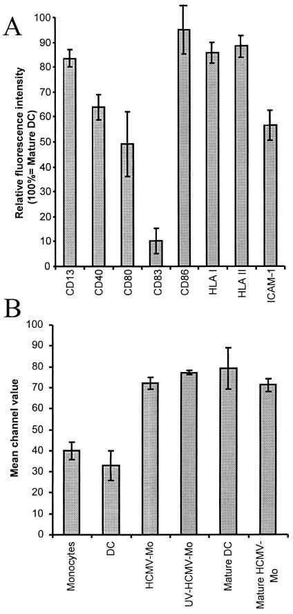
FIG. 7.
HCMV-infected monocytes express high levels of the costimulatory molecule CD86 and an impaired ability to mature in response to LPS treatment. (A) To examine whether the HCMV-infected monocytes (HCMV-Mo) could mature in response to activation by LPS, we stimulated HCMV-infected monocytes and mock-infected dendritic cells (DC) with LPS for 48 h as described in Materials and Methods. The relative fluorescence intensity of the cell surface molecules CD13, CD40, CD80, CD83, CD86, HLA classes I and II, and ICAM-1 was analyzed by flow cytometry. The relative fluorescence intensity of surface molecules on HCMV-infected monocytes following LPS stimulation is shown. Means and standard error of the mean values were calculated from five separate experiments. Relative fluorescence intensity was calculated as (mean fluorescent intensity on “mature” HCMV-infected monocytes/mean fluorescent intensity on mature dendritic cells) × 100. (B) Mean fluorescence intensity values obtained for the costimulatory molecule CD86 on monocytes, dendritic cells, HCMV-infected monocytes, UV-irradiated HCMV-infected monocytes, mature dendritic cells, and mature HCMV-infected monocytes obtained by flow cytometry. The results represent mean and standard error of the mean values obtained from five experiments. (B) We did not observe a difference in cell viability between dendritic cells and HCMV-infected monocytes, as demonstrated by trypan blue dye staining (data not shown), which suggests that the inability of the infected cells to upregulate cell surface molecules was not due to a difference in cell viability.

